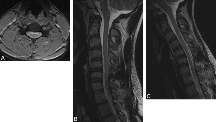
Fig 2.
A 20-year-old man with HD. A, Neutral axial gradient-echo image at the C5 level demonstrates subtle bilateral LOA along the lateral aspects of the lamina bilaterally and spinal cord atrophy, asymmetric to the right. B, Neutral sagittal T2-weighted image also localizes this atrophy to C5-C6. C, Flexion sagittal T2-weighted image demonstrates 2 mm of anterior dural shift. The posterior subarachnoid space is not completely obliterated, and there is no direct spinal cord compression.

