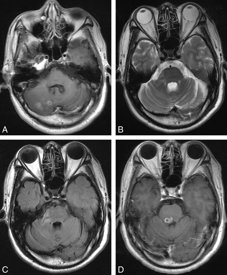
Fig 4.
Case 5. Axial images of a 36-year-old male patient with seizures and left hemiparesis for 17 years. A, Axial postcontrast T1-weighted image shows a lesion with multi-ringlike enhancement in the right cerebral hemisphere. B and C, Four months later, an abnormal branchlike hypointensity is detected in the right cerebellar peduncle on T2-weighted and FLAIR images. D, Eleven months later, a migrated lesion with small ringlike enhancement is detected in the pons. Note that the signal change on B and C indicates the continuity between the primary lesion and the migrated one.

