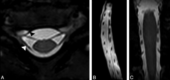
Fig 2.
A, Axial high-resolution MR imaging in a 6-month-old girl with clinically suspected right-sided brachial plexus palsy showing intact right ventral (arrowhead) and dorsal nerve roots on both sides. Compare with normal left-sided nerve roots. Sagittal (B) and (C) coronal reformatted images from the axial data again show normal ventral and dorsal nerve roots at each vertebral level.

