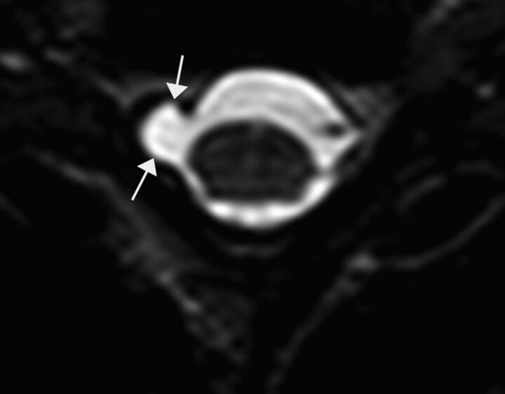
Fig 4.
Axial high-resolution MR imaging in a 4-month-old boy with clinically suspected right-sided brachial plexus palsy shows a pseudomeningocele at right C5–6 level (arrow). Note absent nerve roots on right side suggestive of nerve root avulsion injury. Compare with normal ventral and dorsal nerve roots on the left side.

