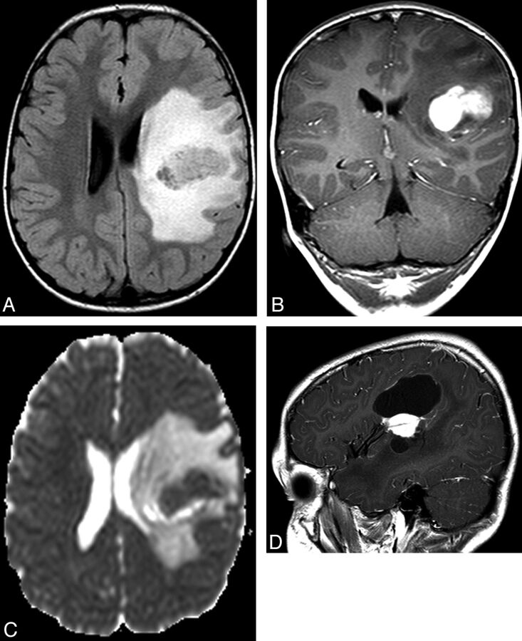
Fig 2.
Left frontal PXA in a 4.8-year-old boy (patient number 2). A, Preoperative axial FLAIR image shows marked vasogenic edema. B, Preoperative coronal T1-weighted gadolinium-enhanced image shows intense enhancement of the lesion. C, The calculated preoperative ADC ratio was 1.06. D, The mass could not be resected completely because of its proximity to the left middle cerebral artery and showed progression over 4 years. Sagittal gadolinium-enhanced T1-weighted image shows the signal void of a middle cerebral artery branch within the enhancing mass.

