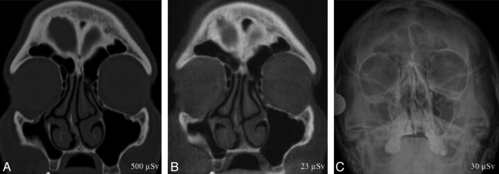
Fig 1.
A 50-year-old woman with vague facial pain for several months and enophthalmos on physical examination. CT (A) and conebeam CT (B) coronal images show inward retraction of the right maxillary walls and apposition of the right uncinate process into the orbital floor, occluding the maxillary sinus infundibulum. The clinical and CT imaging findings were characteristic of silent sinus syndrome. CT scans delineate anatomic structures and demonstrate disease not shown on the x-ray (C). X-ray shows a small right maxillary sinus but is unable to help in the diagnostic work-up of the patient.

