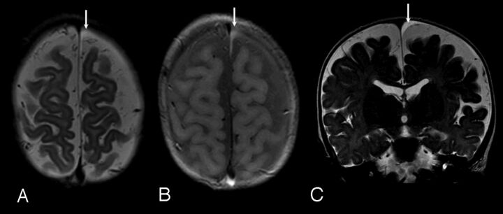
Fig 1.
Case 3. A, Axial FSE T2-weighted image. B, Axial proton density–weighted image. C, Coronal FSE T2-weighted image; 8-month-old girl. Clinical indicatin for examination: macrocrania. Typical small homogeneous subdural collection, similar to those identified in cases 2, 5, and 6. Note diffuse prominence of subarachnoid spaces. Small left frontal vertex subdural collection is identified (arrows), slightly hyperintense to CSF on T2-weighted images (A and C), and moderately hyperintense to CSF on proton density images (B). The collection was isointense to CSF on T1-weighted images and showed no blooming on gradient-echo sequences. Follow-up CT 3 months later showed decrease in prominence of the subarachnoid space, normal ventricles, and no evidence of subdural collection.

