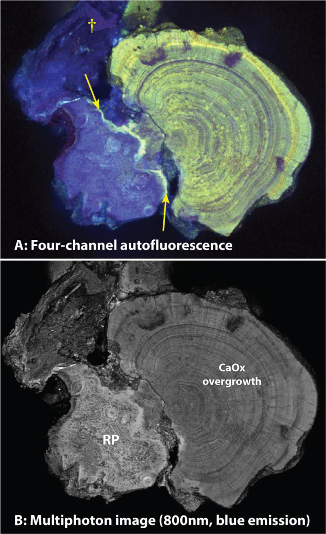
Figure 6.

Fluorescence imaging of stone polished to expose the interface between Randall’s plaque (RP) and the calcium oxalate overgrowth (A, four-channel single-photon, and B, multiphoton microscopy in only the blue range). Note fine detail visible in the multiphoton image (B). Arrows point to a layer of bright fluorescence that indicates enrichment in organic material, perhaps where RP was exposed to urine. Dagger (†) marks a piece of additional RP that apparently was torn up from the tissue during the removal of the stone from the renal papilla and which then adhered to the side of the stone.
