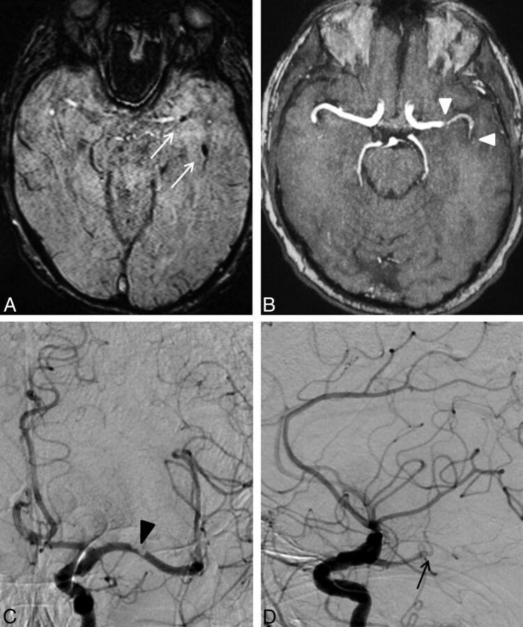
Fig 1.
An 85-year-old man with right-sided hemiparesis and aphasia (NIHSS 13). Pretreatment SWI shows susceptibility vessel signs in the left M1 and M2 segments (A, white arrows). On TOF-MRA (MIP), diminished flow signal is seen distal to the thrombus fragment in the M1 segment with complete loss of flow signal at the site of the distal thrombus fragment (B, white arrowheads). DSA demonstrates incomplete occlusion of the vessel lumen by a thrombus in the M1 segment (C, anteroposterior projection, black arrowhead). On the lateral projection, occlusion of the temporo-occipital M2 branch by a more distal thrombus fragment is visible (D, black arrow). Although both TOF-MRA and DSA can show the distal vessel occlusion, only SWI allows estimation of the length of the distal fragment and exclusion of additional distal thrombi.

