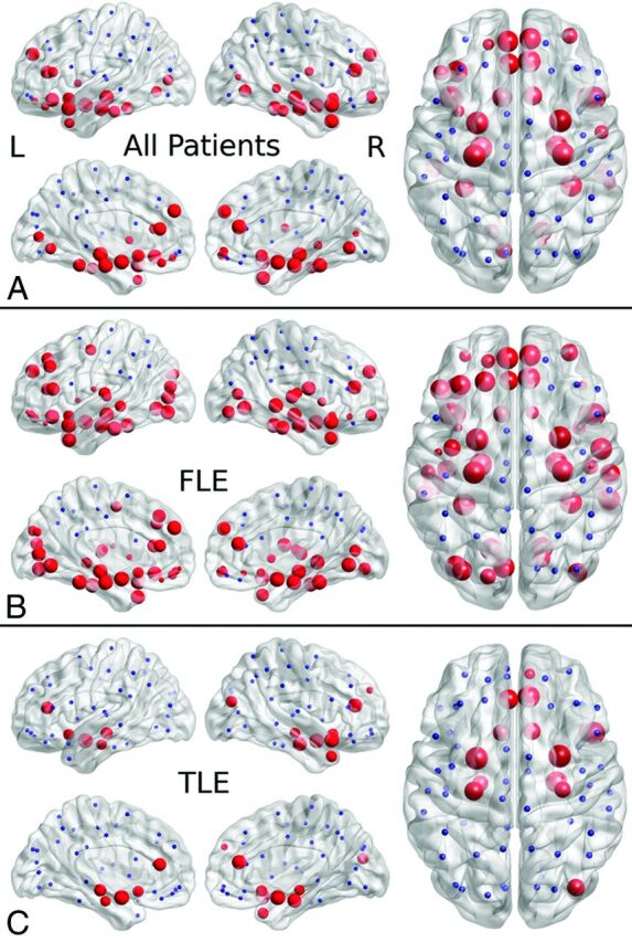
Fig 1.

Reduced nodal efficiency in localization-related epilepsy (A), frontal lobe epilepsy (B), and temporal lobe epilepsy (C) in patients relative to controls. The disrupted nodes with reduced nodal efficiency are shown in red, and the unaffected nodes are shown in blue. The size of the nodes is related to the significance of between-group differences in nodal efficiency, with larger nodes representing more significant (lower P value) reduced nodal efficiency in patients relative to controls.
