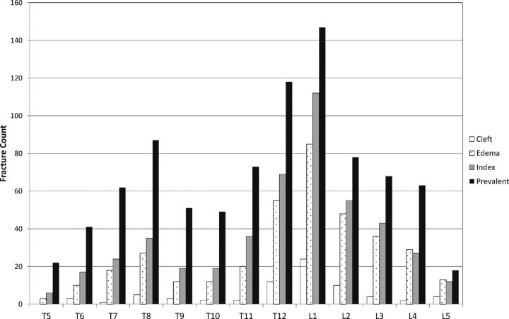
Fig 2.
Distribution of index and prevalent fractures and those with edema and vacuum cleft for the BKP and VP groups combined. Index levels (those identified as treatment levels) and prevalent fractures (all radiographic fractures assessed by the core laboratory) are shown, identified from standing lateral x-ray films with 379 of 381 treated patients contributing data. The distribution of levels with edema and those with vacuum cleft is shown on the basis of available MR imaging at baseline (294 of 381 treated patients).

