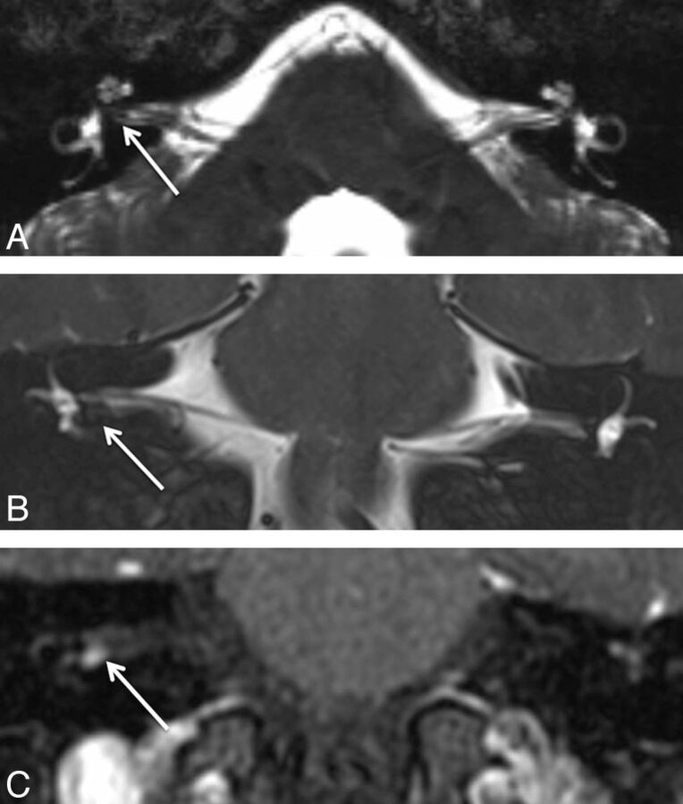
Fig 1.
A 29-year-old man with hearing loss on the right. A, Axial CISS image shows subtle asymmetric hypointense signal within the fundus of the right IAC (arrow), which could be dismissed as volume averaging. B, Coronal T2WI better demonstrates the hypointense lesion (arrow) along the inferior right IAC fundus, which is confirmed to be a 4-mm enhancing mass (arrow) on postcontrast coronal T1WI (C).

