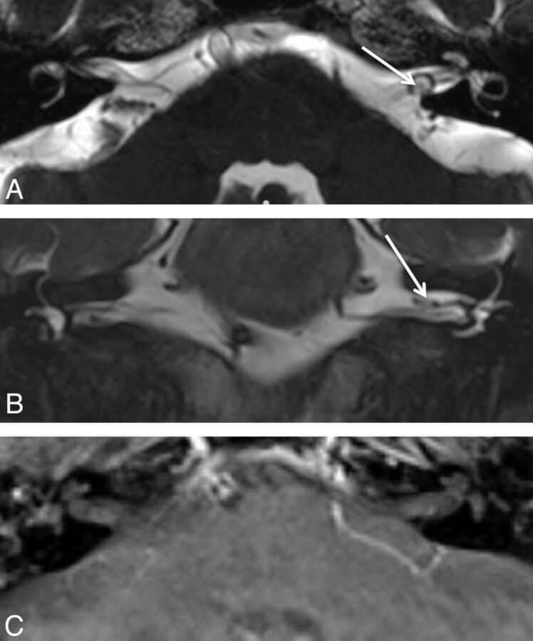
Fig 3.
A 71-year-old man with vertigo. A, Axial CISS image demonstrates a hypointense focus surrounded by a loop of the anterior inferior cerebellar artery, which was thought to represent a small IAC lesion. B, Coronal T2WI shows the same hypointense focus. C, Postcontrast axial T1WI shows no enhancing lesion, indicating that the hypointense focus on the screening study was volume averaging related to the adjacent artery.

