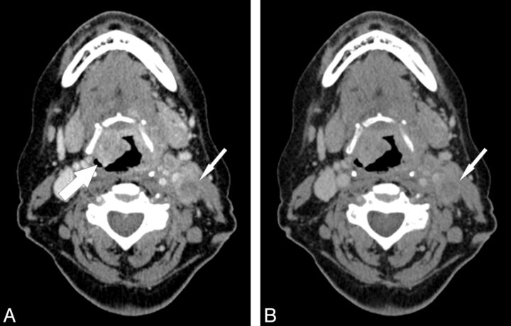
Fig 1.
A 61-year-old female patient with a primary supraglottic laryngeal carcinoma (T4a N2c). Low-tube-voltage acquisition (A) improves tumor attenuation (large arrow) compared with the standard blended 120-kVp image series (B) and also shows a higher contrast and improved depiction of cervical lymph node metastasis (small arrows) (window settings: width, 400 HU; level, 80 HU).

