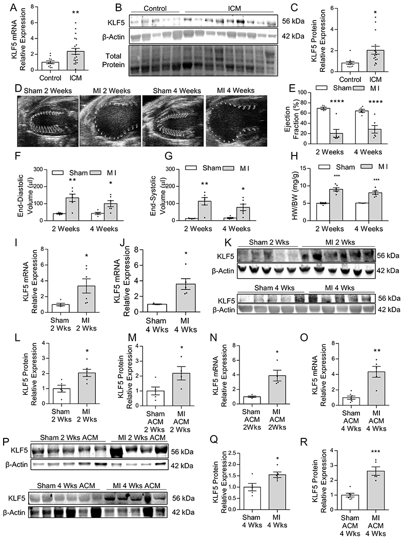
Figure 1: Cardiomyocyte KLF5 is Increased in Ischemic Heart Failure –

KLF5 mRNA (A), Western blotting (B) and quantification (C) in heart tissue obtained from healthy control and end-stage ischemic heart failure patients. n=11 control, n=20 ICM patients for mRNA analysis; n=5 control and n=9 ICM patients for Western Blotting analysis. *p<0.01 by Welch’s t-test. Representative parasternal long-axis images of the ventricle with wall motion shown in the traced area (D), and measurements (VevoStrain software) of ejection fraction (E), end-diastolic volume (F), and end-systolic volume (G) in sham and MI C57Bl/6 mice 2-weeks and 4-weeks post-surgery. Heart weight normalized to body weight (H). n=5 sham 2-weeks and 4-weeks, n=6 MI 2-weeks and 4-weeks. *p<0.05, **p<0.01, ****p<0.0001; panel E and H analyzed using t-test, panel F and G analyzed using Welch’s t-test. Expression of KLF5 mRNA in heart tissue at 2-weeks (I) and 4-weeks (J) after MI. n=5 sham 2-weeks and 4-weeks, n=6 MI 2-weeks and 4-weeks *p<0.05 by Welch’s t-test. Western blotting (K) with densitometric quantification of KLF5 protein levels in whole heart tissue from C57BL/6 mice 2-weeks (L) and 4-weeks (M) post MI. n=5 sham 2-weeks and 4-weeks, n=5–6 MI 2-weeks and 4-weeks. *p<0.05 by t-test. KLF5 mRNA in isolated adult cardiomyocytes (ACM) 2-weeks (N), and 4-weeks (O) after MI. n=4–6 sham ACM 2-weeks and 4-weeks, n=4–5 MI 2-weeks and 4-weeks. *p<0.05, **p<0.01 by Welch’s t-test. Western blotting (P) with densitometric quantification of KLF5 protein levels in isolated adult cardiomyocytes from C57BL/6 mice 2-weeks (Q) and 4-weeks (R) post-MI. n=5–6 sham ACM 2-weeks and 4-weeks, n=4–5 MI 2-weeks and 4-weeks. *p<0.05, ***p<0.001 by t-test.
