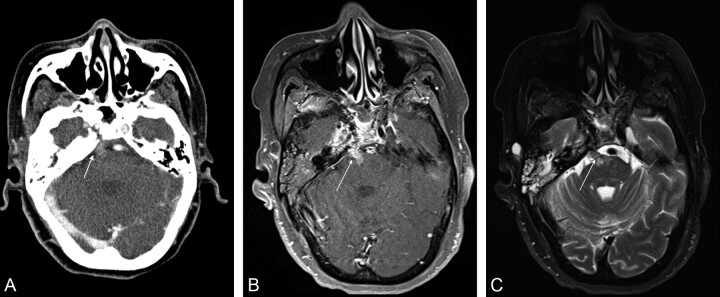
Fig 1.
Axial CT and MR images in a 61-year-old woman treated previously for nasopharyngeal carcinoma. A, Contrast-enhanced CT image shows abnormal thickening and mild enhancement along the right sixth cranial nerve (arrow). B, T1-weighted fat-saturated gadolinium-enhanced axial image through the level of the pons more clearly demonstrates the band of abnormal enhancement and thickening extending from the clivus into the right aspect of the pons (arrow). C, Fat-saturated axial T2-weighted image also shows the lesion to be of heterogeneous internal signal intensity with clear extension into the pons (arrow).

