Figure.
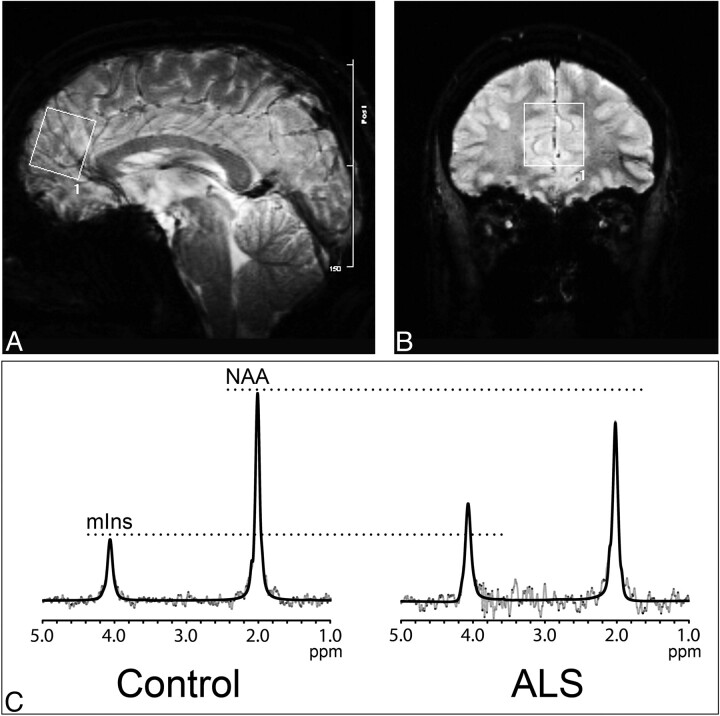
Sagittal (A) and coronal (B) gradient-weighted MR images demonstrate voxel placement in the mesial prefrontal cortex. Representative spectra (C, gray lines) and LCModel fit (C, dark lines) with peaks identified for mIns and NAA are shown for a control subject (left) and patient with ALS (right); note the increased mIns and decreased NAA in the patient with ALS compared with the control subject.

