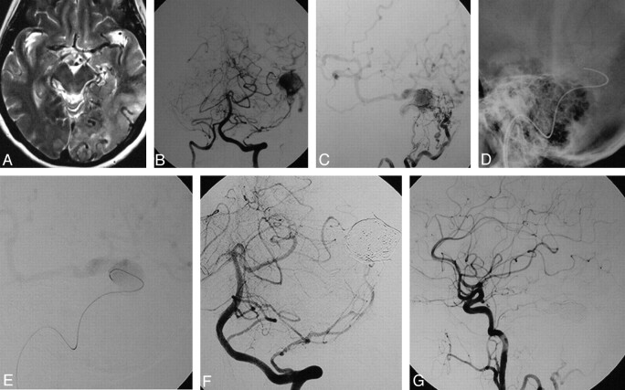
Fig 2.
DAVF into a left “isolated” transverse sinus with cortical venous reflux. This 59-year-old female patient presented with alteration of consciousness and vertigo. A, Axial T2-weighted image shows dilation of cortical veins and hypersignal of the brain parenchyma as a sign of venous congestion in the left temporal and occipital lobes. B and C, Vertebral artery injections in an AP plane (B) and lateral occipital artery injections (C) demonstrate a DAVF at the left transverse sinus with an isolated enlarged venous pouch and cortical venous reflux with supply from multiple small branches of the occipital arteries and a dural branch of the left vertebral artery. D, With the guiding catheter in the left jugular bulb, a 0.035-inch guidewire is advanced through the occluded sigmoid sinus into the venous pouch. E, The track is used by a microcatheter, and the position of the microcatheter is checked with a careful injection once in the venous pouch. F and G, Following packing of the sinus with coils, control angiograms of the left vertebral artery (F) and the left common carotid artery (G) demonstrate complete obliterations of the DAVF.

