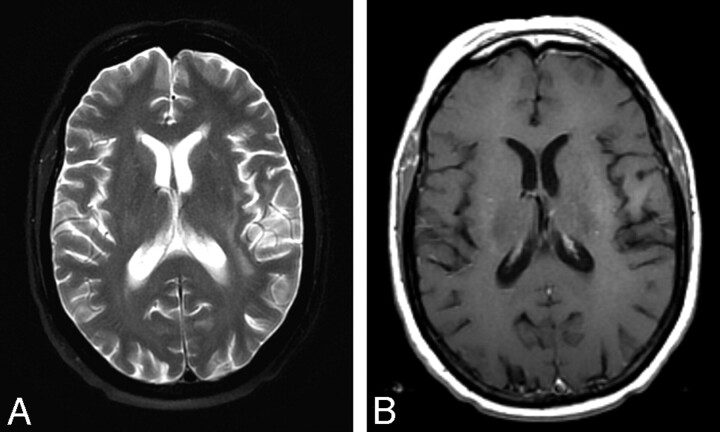
Fig 3.
The patient was admitted and received chemotherapy with methotrexate, vincristine, and rituximab. Follow-up MR imaging was performed 2 cycles into her chemotherapy. A, T2-weighted axial FSE image shows only residual edema as high T2 signal intensity in the deep white matter of the left frontal parietal lobes. B, Enhanced T1-weighted axial spin-echo image shows resolution of the enhancing mass.

