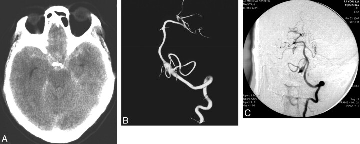
Fig 2.
Images obtained in a 59-year-old man who presented with SAH due to a ruptured right VA-PICA aneurysm. A, CT scan. B, 3D reconstruction of the right VA injection. The patient had undergone right VA coil occlusion, after which he recovered. C, Left VA angiogram after right VA occlusion reveals that the right PICA is filled via the left VA and the aneurysm is decreased in size.

