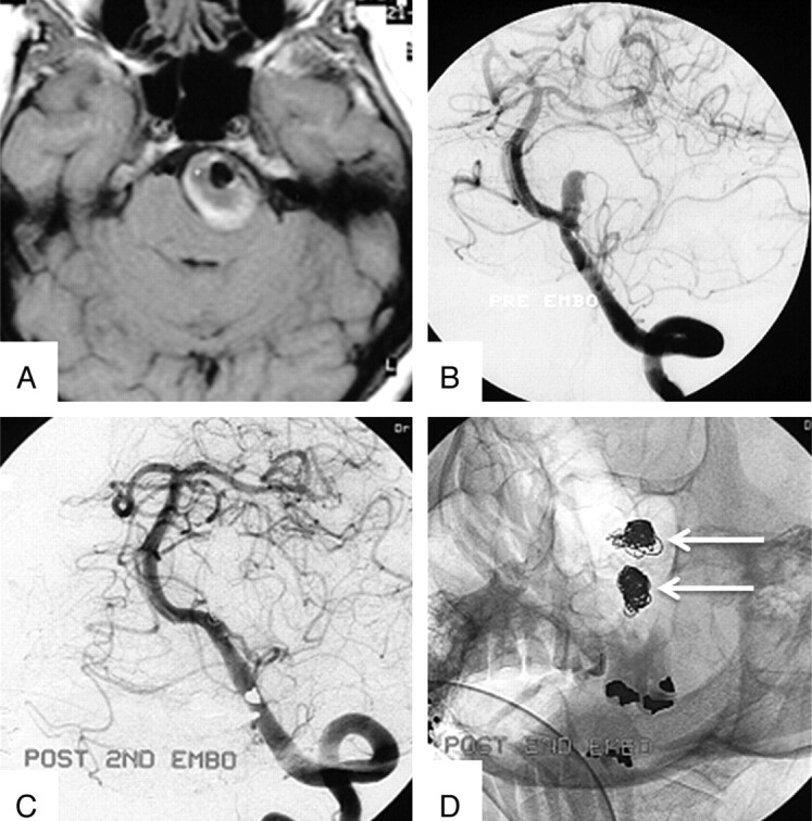
Fig 5.

A 55-year-old woman with brain stem compression from a partially thrombosed PICA aneurysm. A, Axial T1-weighted MR image at presentation shows a 32-mm left PICA aneurysm with a large intraluminal thrombus. B, Left vertebral angiogram shows the small aneurysm lumen. C and D, Angiogram (C) and nonsubtracted image (D) after the second coiling show 2 separate coil meshes: The first coil mesh has migrated into the thrombus, and the second coil mesh occludes the lumen.
