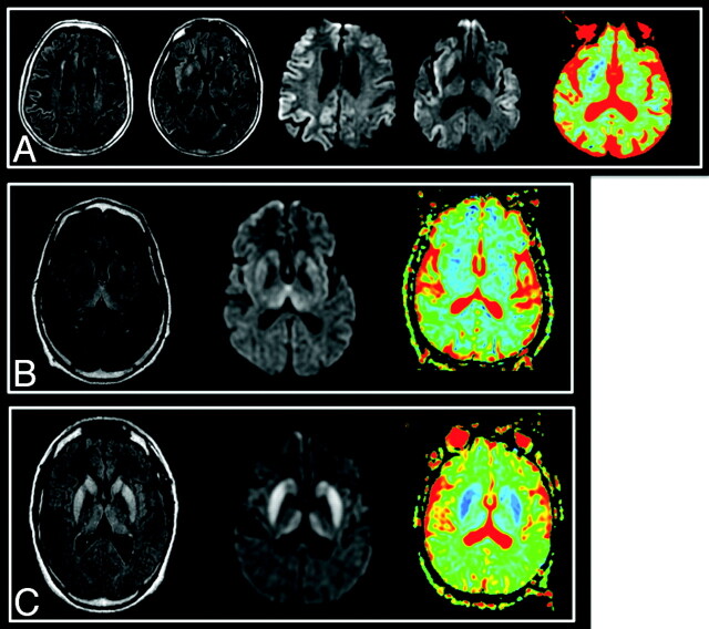
Fig 2.
Typical images in cases of sCJD (A), vCJD (B), and gCJD (C). FLAIR, DWI, and ADC map, respectively, are shown. Areas of increased signal intensity, which involve the cortex and the striatum are more extensive and more clearly visible on diffusion images. On the basal ganglia, these changes are associated with a decreased ADC. There is widespread involvement of the cortex in the patient with sCJD. gCJD and vCJD both present with lesions of the thalamus and lenticular nuclei. However, in the variant case, as opposed to the genetic one, the areas of high signal intensity are more pronounced in the pulvinar than in the striatum as has been previously described in this phenotype.

