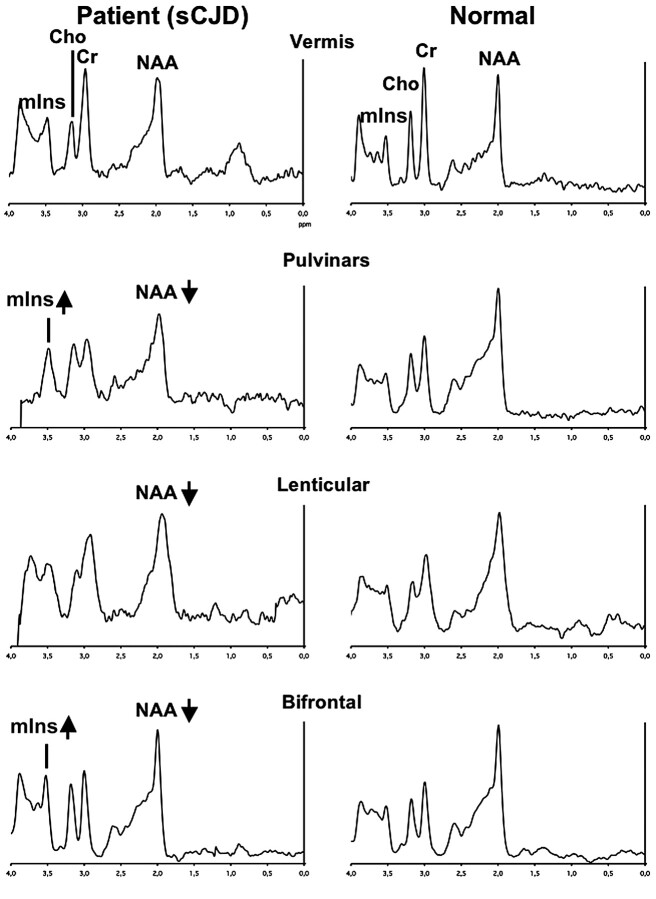
Fig 3.
Typical spectra recorded in a patient with sCJD (left column) and in a healthy volunteer (right column) in the vermis, pulvinar, right lenticular nucleus, and frontal gray matter. Metabolic anomalies are observed on the bifrontal voxel (decreased NAA and increased mIns), on the lenticular voxel (decreased NAA), and on the bipulvinar voxel (decreased NAA and increased mIns). Note that to get an accurate idea of NAA and mIns variations, one should compare their resonances with “stable” metabolites (eg, Cr).

