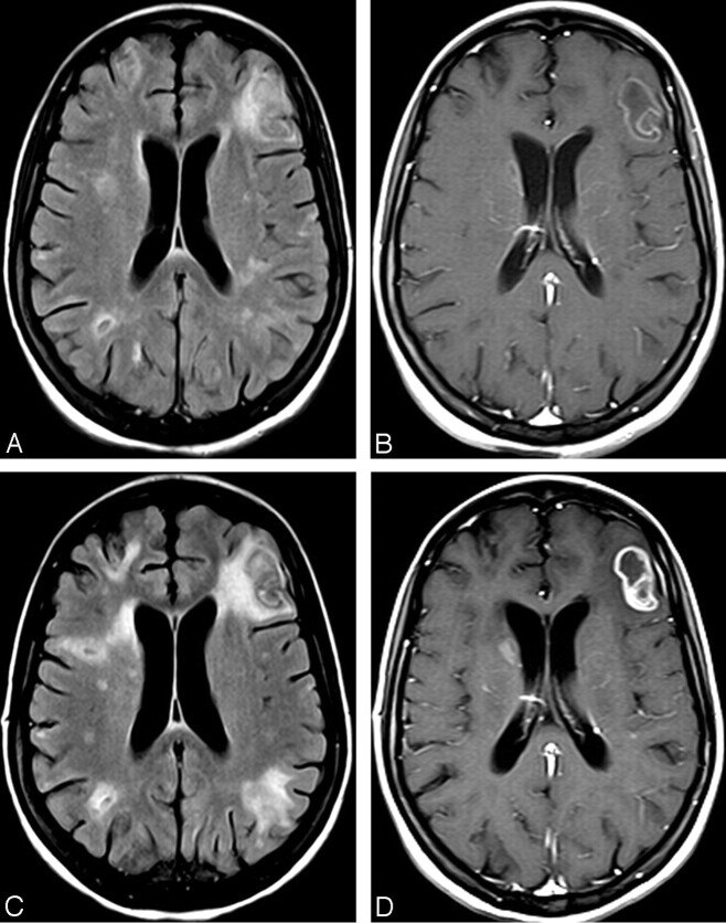
Fig 1.
A, FLAIR image at admission shows multiple areas of high signal intensity, some with a central area of low signal intensity, located in the subcortical and periventricular white matter. B, T1-weighted image after gadolinium administration demonstrates mild ring enhancement in the left frontal lesion, as well as in the lesion in the head of the right caudate nucleus. C, FLAIR image 1 month later shows significant increase in the size of the areas of high signal intensity in the white matter. D, T1-weighted image after gadolinium administration demonstrates stronger enhancement in the lesions at the left frontal lobe and right caudate nucleus, as well as new nodular areas of enhancement bilaterally.

