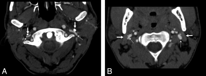
Fig 1.
Examples of extrinsic compression by the styloid process and the posterior belly of the digastric muscle. A, In this axial CT image, there is bilateral compression of the internal jugular veins (dashed arrows) by the styloid processes (solid arrows). The vein takes a pancake-like configuration at this level. B, Similar extrinsic compression, caused by the posterior belly of the digastric muscle (solid arrows) of both internal jugular veins (dashed arrows), though to a lesser degree. In both cases, the vein is compressed anterolaterally by the described structure (styloid process or digastric muscle). Along the medial and posteromedial aspect of the vein, the adjacent vertebra is the most rigid structure and will provide the other side of the extrinsic compressive “pincer.”

