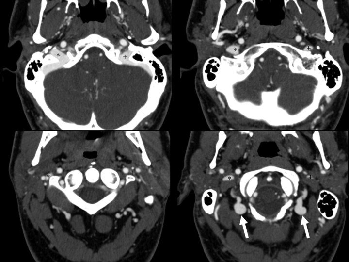
Fig 2.
Example of bilateral collateral vein filling. Four axial images from a CTA of the neck demonstrate bilateral posterior condylar and upper deep cervical vein filling (arrows). Note how contrast in these veins is isoattenuated with the jugular bulb. The left (+) and right (*) internal jugular veins are marked.

