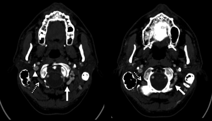
Fig 3.
Examples of condylar veins with and without collateral flow. Two axial images from a CTA of the neck demonstrate differential contrast attenuation in the condylar veins. The left condylar veins (solid arrows) are isoattenuated with the right internal jugular vein (arrowhead). The left internal jugular vein is markedly attenuated. Note that the right-sided condylar veins (dashed arrows) have similar caliber to the left but are not filled with contrast of the same attenuation. This would suggest that the venous drainage of the brain in this patient does use the left condylar veins but not the right.

