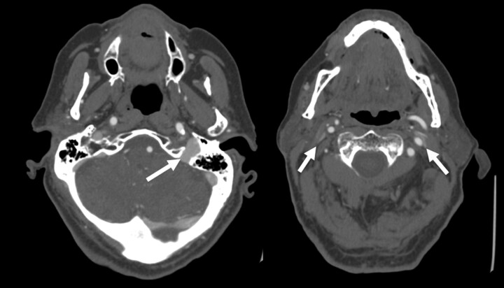
Fig 4.
Example of severe bilateral jugular venous stenosis without collateral formation. Two axial images from a CTA of the neck demonstrate a dominant left internal jugular vein (arrow, left image). The more caudal image (right) shows severe pancake-like flattening of the internal jugular veins bilaterally (arrows) but with no condylar collateral filling seen.

