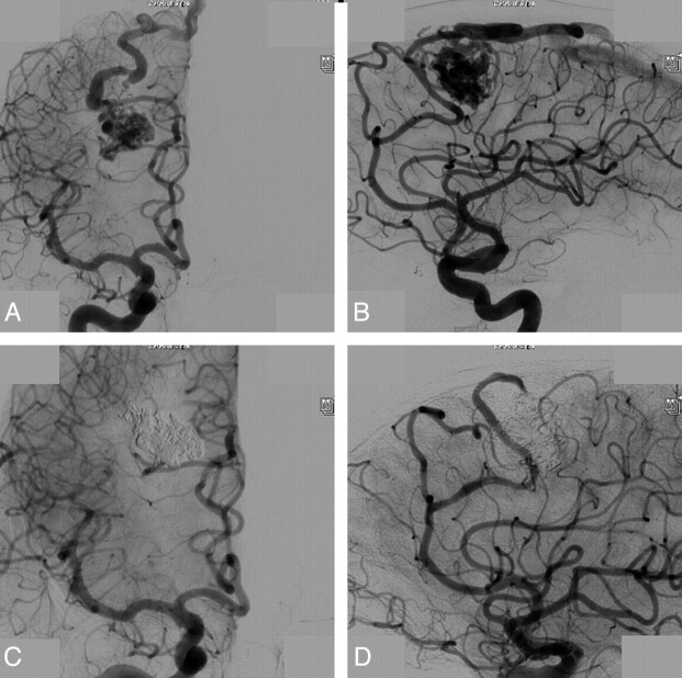
Fig 2.
A 53-year-old man with seizures from a small frontal AVM. Frontal (A) and lateral (B) views from a right internal carotid angiogram demonstrate a small frontal AVM with 2 feeders from the anterior cerebral artery, a well-delineated nidus, and a single draining vein. C and D, Complete obliteration after embolization with Onyx.

