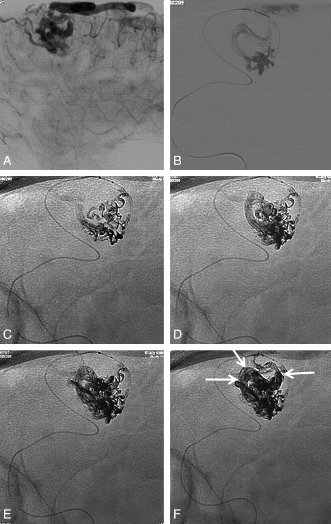
Fig 3.

Embolization technique with Onyx of a small frontal AVM (same patient as in Fig 2). A, Late phase of the lateral view of an internal carotid artery angiogram shows the dominant draining vein. B, Superselective angiogram shows the lower part of the nidus and the origin of the draining vein. C–F, Different stages of a 64-minute Onyx injection show progressive filling of the nidus, including the dominant draining vein (arrows in F).
