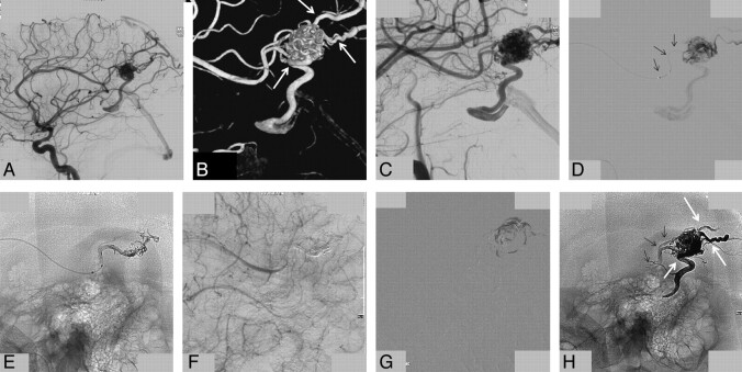
Fig 4.
A 33-year-old man with a ruptured small parietal AVM. A and B, Lateral view of a right internal carotid angiogram and off-lateral 3D rotational image demonstrate a small parietal AVM supplied by the pericallosal artery with 1 deep and 2 superficial draining veins (arrows in B). C, Off-lateral magnification angiogram, identical to the 3D projection in B, best shows the proximal draining veins emerging from the nidus. D, Superselective angiogram through the microcatheter demonstrates a 3-cm distal end allowing reflux of Onyx (arrows). Early phase of Onyx injection shows a 2-cm reflux with little penetration into the nidus (E), but the angiogram suggests complete occlusion (F). G, Subtracted fluoroscopic image demonstrates progressive nidus filling with Onyx after apparent complete angiographic obliteration. H, Final Onyx cast with a 2-cm reflux in the feeder (small arrows) and occlusion of all 3 draining veins (arrows, compare with B).

