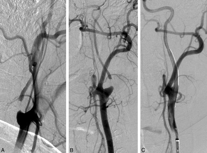
Fig 1.
Left common carotid artery angiography showing pseudo-occlusion of internal carotid artery. Oblique view (A) and lateral view (B). The internal carotid artery filling was delayed regarding to the external carotid artery. C, Immediate control after stent deployment (7 × 40 mm Carotid Wallstent) and postdilatation with a 4 × 20 mm balloon. No predilation was performed.

