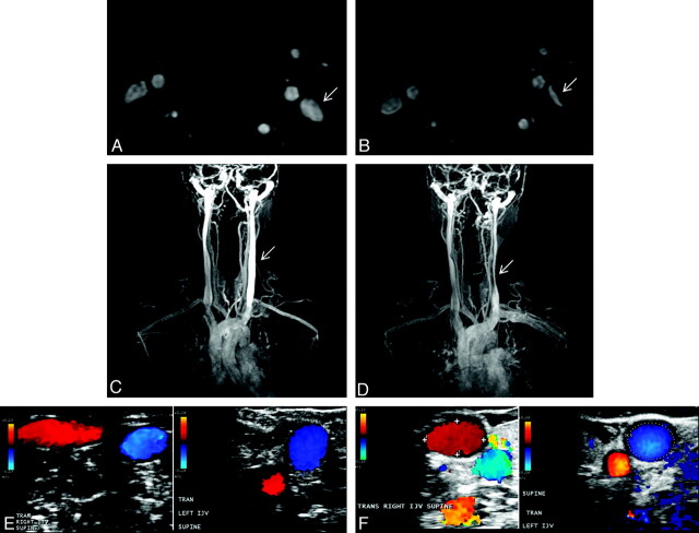
Fig 3.
Variability between the baseline (A, C, and E, TOF, TRICKS, and Doppler sonography, respectively) and follow-up (B, D, and F, TOF, TRICKS, and Doppler sonography, respectively) examinations in a 42-year-old healthy female control. Flattening of the left IJV (arrows) at follow-up is noted on the TOF (B) and TRICKS (D), whereas Doppler sonography shows normal examination findings like those at baseline (F).

