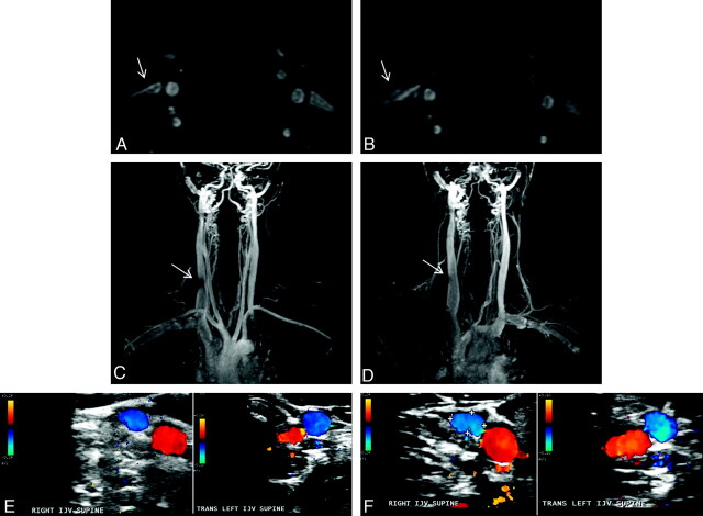
Fig 4.
Variability between the baseline (A, C, and E, TOF, TRICKS, and Doppler sonography, respectively) and follow-up (B, D, and F, TOF, TRICKS and Doppler sonography, respectively) examinations in a 39-year-old healthy male control. Flattening of the right IJV (arrows) present at the baseline (A and C) examination is not present at follow-up (B and D). Doppler sonography examination shows normal findings at baseline (E) and follow-up (F).

