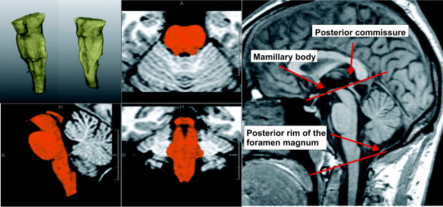
Fig 1.
Example of quantitative brain stem volumetry. Upper left: 3D segmentation of the brain stem. Lower left and middle: brain stem segmentation in orthogonal views. The borders of the brain stem are defined according to the method proposed by Luft et al.18 Right: midsagittal view showing anatomic landmarks.

