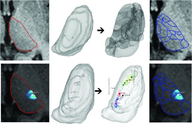
Fig 1.
Atlas-to-patient registration by the outline-based SSM and the hybrid SSM method. First row: alignment of atlas data (image 3) to the subject's gross, visible thalamus outlines through a statistical shape model–based registration (image 1, red outlines: thalamus borders; image 2: 3D visualization). Second row: atlas-to-patient registration by using the hybrid SSM method. Matching is guided by the weighted contribution of the gross visible thalamus borders and the DTI-based intrathalamic markers of connectivities (panel 1: visible outlines and the center-of-mass point of the connectivity map to the postcentral gyrus in panel 2; panel 3: alignment of thalamus outlines and DTI markers). The resulting thalamus maps are given in the rightmost panel. Abbreviations of the connectivity-based landmarks are given according to the cortical target areas used. FP indicates frontal pole; MFG, middle frontal gyrus; SFG, superior frontal gyrus; CDN, caudate nucleus; SMC, supplementary motor cortex; PREC, precentral gyrus; PSC, postcentral gyrus; SPL, superior parietal lobule; CEREB, cerebellum; LOCS, lateral occipital cortex, superior division.

