
Fig 1.
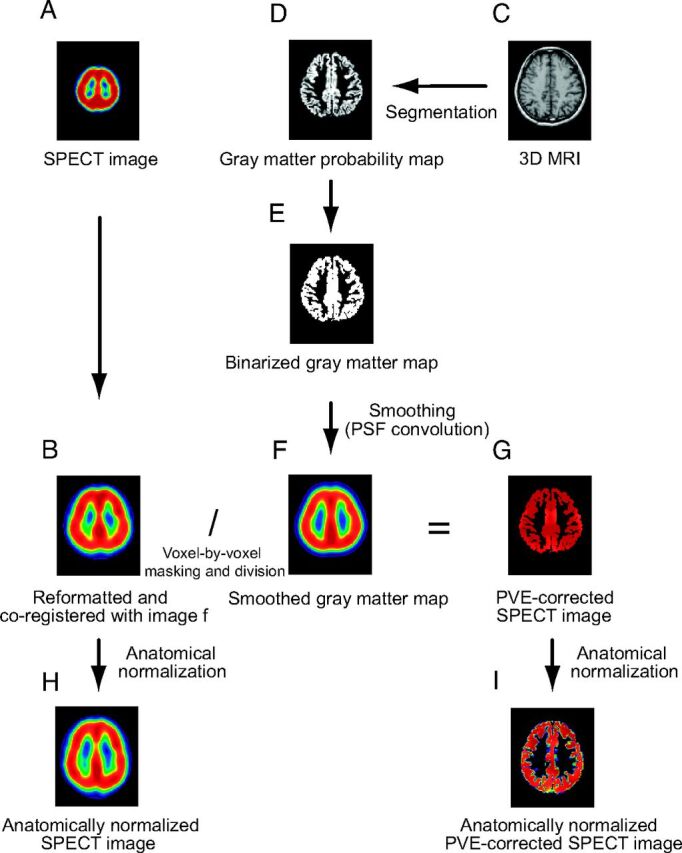
A, I-123 iomazenil SPECT image. B, Automatic coregistration of the I-123 SPECT image with the MR image. The maps are simultaneously reformatted to a matrix that is the same size as the referenced smoothed gray matter map. C, 3D MR image obtained before surgery. D, The MR image is segmented into a gray matter map. E, The gray matter probability map is subsequently binarized. F, Binary map for gray matter convoluted with the point-spread function (smoothed gray matter map). G, Smoothed gray matter map masked by the image E. The coregistered I-123 SPECT image is divided by using the masked smoothed gray matter map on a voxel-by-voxel basis. H, Image B anatomically normalized by the spatial normalization matrices generated in the segmentation process. I, Image G anatomically normalized in the same manner as image H.