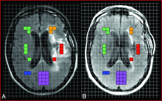
Fig 1.
Representative example of voxel placement onto registered grid overlay on the pre-SIACI (A) and post-SIACI (B) of bevacizumab T2-weighted FLAIR MRS localizer images demonstrating the 5 aforementioned regions of interest selected for analysis: red = enhancing component; orange = nonenhancing T2-hyperintense signal abnormality; green = matched contralateral “normal” parenchyma (corresponding to cumulative area of enhancing and nonenhancing voxels); blue = normal contralateral white matter; purple = normal cortex. ROIs were selected on the pretreatment T2-weighted FLAIR MRS (A) and these 5 identical anatomic areas were then carried over and plotted on the posttreatment T2-weighted FLAIR MRS (B).

