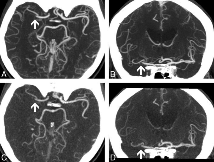
Fig 1.
A and B, CTA, transversal and coronal MIP reconstructions; image thickness 10 mm. C and D, VPCTA, transversal and coronal MIP reconstructions; image thickness 10 mm. The same window settings were used for all reconstructions. Occlusion of the distal main branch of the right middle cerebral artery is demonstrated in CTA and VPCTA (arrows). In VPCTA, veins are less contrasted.

