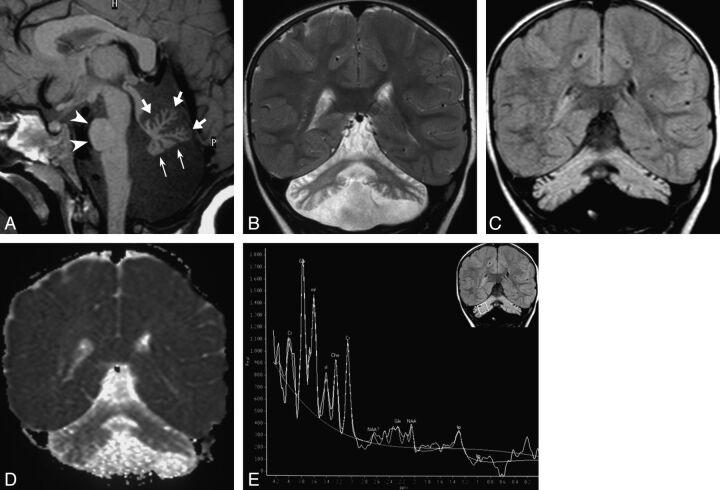
Fig 1.
Typical MR imaging findings in CDG-1a; case 4 at age 2 years. A, Sagittal T1-weighted image shows shrunken appearance of the vermis, with commensurate folial volume loss and fissural enlargement involving the anterior lobe and declive (thick arrows) and flattened inferior vermis (thin arrows). The pontine protuberance is small (arrowheads). B, Coronal T2-weighted and (C) FLAIR images show shrunken, hyperintense hemispheric cerebellar cortex bilaterally; the cerebellar white matter is also mildly hyperintense compared with supratentorial white matter, resulting in a “bright cerebellum” appearance. D, Coronal ADC map shows the cerebellum is hyperintense compared with the supratentorial brain, consistent with facilitated diffusion. E, 1H-MRS findings; single-voxel PRESS technique at TE 23 ms. Upper right coronal FLAIR image shows placement of a 18 × 18 × 18 mm voxel on right cerebellar hemisphere. Corresponding spectrum shows markedly reduced NAA peak, mildly reduced Cho, and elevated inositol (mI and sI) resonances. There is a high alpha-Glx peak at 3.8 ppm, perhaps in relation to hepatic dysfunction in this patient. Glx indicates glutamine/glutamate complex.

