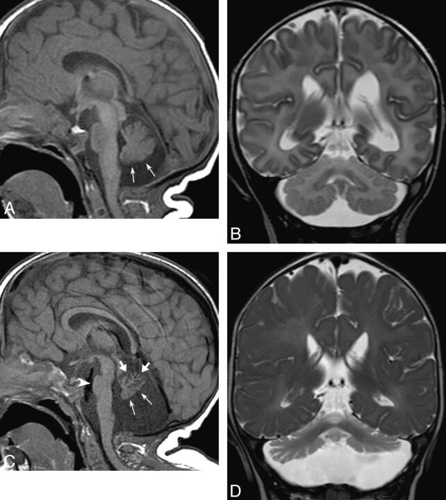
Fig 2.
Evolution of findings in CDG-1a; case 1 at age 12 days (A and B) and 11 months (C and D). At presentation, sagittal T1-weighted image (A) shows mild hypoplasia of the inferior vermis (thin arrows); the pons and superior vermis appear normal. Coronal T2-weighted image (B) shows normal cerebellar hemispheres. At 11-month follow-up, sagittal T1-weighted image (C) shows considerable volume loss of the vermis. Notice, in particular, atrophic involution of the anterior lobe and declive (thick arrows), with corresponding fissural enlargement, whereas the inferior vermis retains a flattened appearance (thin arrows), with not so large fissures. The pontine protuberance is also diminished in size (arrowhead). Coronal T2-weighted image (D) also shows reduced size of both cerebellar hemispheres. The cerebellar cortex is hyperintense.

