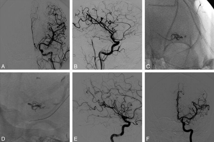
Fig 2.
Case 1. Final angiograms at the end of the procedure in anteroposterior (AP; A) and lateral (B) views. Onyx cast at the end of the procedure (DSA unsubtracted images) in AP (C) and lateral (D) views. Six-month DSA follow-up in AP (E) and lateral (F) projections confirming the complete and persistent occlusion of the AVM.

