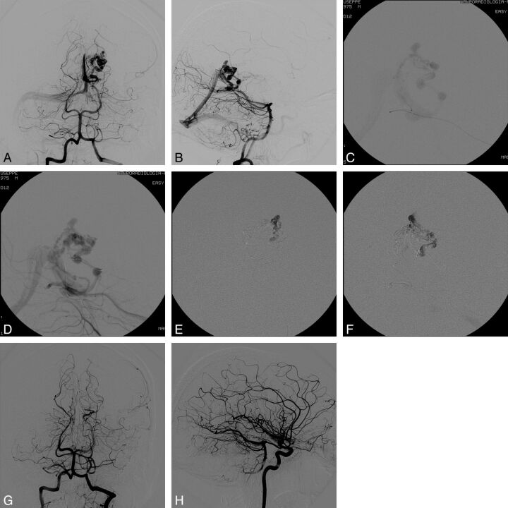
Fig 3.
Case 4. Pretreatment DSA evaluation in anteroposterior (AP; A) and lateral (B) views. Visualization of the nidus and the draining venous system with transvenous injection through the microcatheter positioned at the origin of the draining vein (C) and the intermediate catheter positioned within the straight sinus (D). Intranidal penetration of Onyx through transvenous injection from the main venous drainage (E and F). DSA evaluation at the end of the procedure in AP (G) and lateral (H) views: The AVM is completely obliterated.

