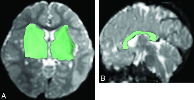
Fig 1.
Regions of interest (green shaded areas) were manually drawn on axial B0 image (A) to include the ipsilateral caudate head, internal capsule, lentiform nucleus, external capsule, and thalamus for reconstructing the PF on one side, and on sagittal B0 image (B) to include the corpus callosum for reconstructing the CF.

