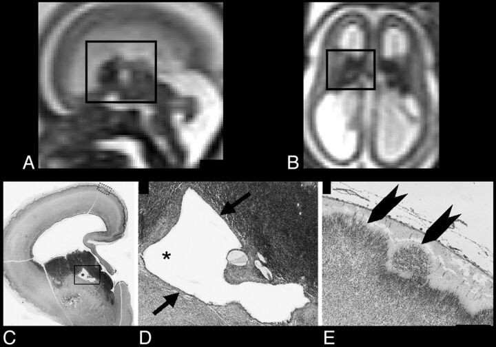
Fig 2.
A and B, Sagittal and axial single-shot FSE T2-weighted sections of case 1 (22-week GA), respectively: black rectangle encompasses GE and relative cavitation. C, Thionin-stained paraffin coronal section shows a hemisphere at the level of GE cavitation (asterisk). D, Higher magnification of the black rectangle area in C, illustrating the regular border (epithelium-like structure) of cavitation (arrows). E, Higher magnification of the cortex (dotted rectangle in C): heterotopic cortical plate neurons extending in the marginal zone (arrowheads). Scale bar = 300 μm.

