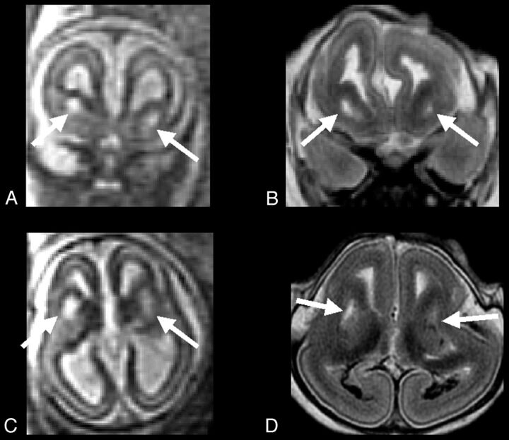
Fig 4.
A and C, Single-shot FSE T2-weighted sections from case 3 (23-week GA) prenatal study, show large symmetric GE region cavitations (arrows). B and D, FSE T2-weighted corresponding MR autopsy sections, which confirm the prenatal findings (arrows): cavitations appear to have regular smooth margin, albeit brain was compressed and deformed during delivery.

