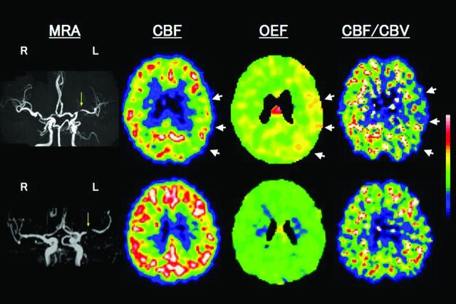
Fig 2.
Upper row shows examples of PET images for a patient with left (L) remote (41 months) symptomatic MCA stenosis with MP. MRA shows severe stenosis of the L MCA. PET study shows reduced CBF, increased OEF, and decreased CBF/CBV in the L hemisphere with MCA stenosis (arrows). A subsequent transient ischemic attack (a transient episode of sensory aphasia) occurred 14 days after the PET study. Lower row shows a patient with L asymptomatic MCA stenosis with normal cerebral hemodynamics.

