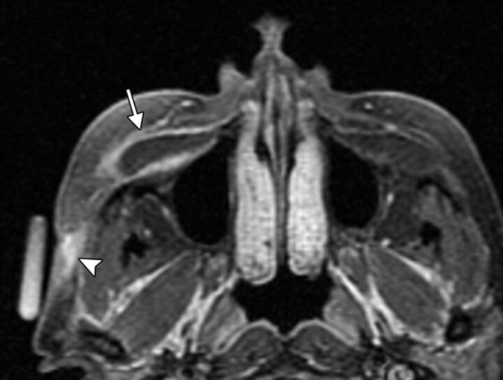
Fig 15.
Implant infection. Axial fat-suppressed T1-weighted MR image shows abnormal enhancement surrounding a fluid-filled right cheek implant pocket (arrow), which extends through the right lateral cheek subcutaneous tissues to an overlying skin defect, representing a draining sinus (arrowhead). An external marker is positioned over the draining sinus.

