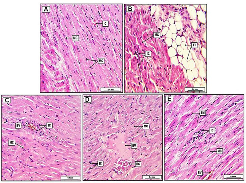
Figure 4.
Photomicrograph of heart from groups; (A) control group, display typically arranged myocardial cells (MC), with their oval nuclei and light pinkish cytoplasm, in addition, the section shows some congested blood capillaries (C). (B) Doxorubicin group (DOX), reveal remarkable fatty infiltration (FI) within the cardiac muscle, together with significant fatty degeneration (FD), the section shows severe degenerative changes in myocardial muscle (MC), together with the infiltration of inflammatory cells (IC). (C) Doxorubicin, quercetin and sitagliptin group (DOX+QC+STN), display slight pre-vascular cuffing of inflammatory cells (IC), together with vascular congestion in some blood vessels (BV), moreover, some myocardial cells (MC) show acidophilic cytoplasm with condensate nuclei. (D) Doxorubicin and quercetin group (DOX+GC), demonstrate slight to moderate infiltration of inflammatory cells (IC), the section reveals some blood vessels (BV) with semi-coagulated red blood cells (BC) and eosinophilic plasma-rich protein. Myocardial muscle (MC) shows oval nuclei with cross-striated appearance. (E) Doxorubicin and sitagliptin group (DOX+STN), reveal slight infiltration of inflammatory cells (IC), the section shows some eosinophilic myocardial muscle cells (EM) with condensate nuclei, however, some other myocardial cells (MC) demonstrate typical cross striation together with their oval nuclei. Some blood vessels appear congested (BV). H&E. Scale bars: 0.4 mm.

