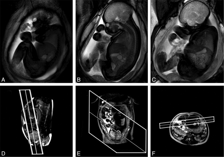
Fig 1.
Example of a cine acquisition at GA 36 weeks. A–C, Successive frames from a multisection cine image sequence (section thickness, 30 mm). D–F, Piloting procedure used to set up the scan. Upper and lower limbs, trunk, and head are all visible within the FOV. D, Fetal localizer scan, which is used to generate sagittal-oriented cine data; E and F, Maternal pilots in orthogonal planes used to ensure that the prescribed sections have FOVs that cover the full extent of maternal tissue.

